|
|
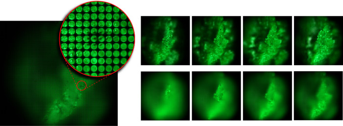
| At left is a light field captured by photographing a speck of fluorescent crayon wax through a microscope objective and microlens array. The objective magnification is 16x, and the field of view is 1.3mm wide. The image consists of 170x170 subimages, one per microlens, each depicting a different part of the specimen. An individual subimage contains 20x20 pixels, each representing a different point on the objective lens and hence a unique direction of view of the specimen. By extracting one pixel from each subimage, we can produce perspective views of the specimen, a sequence of which is shown at top-right. Alternatively, by summing the pixels in each subimage, we can produce orthographic views with a shallow depth of field, like an ordinary microscope but of lower spatial resolution. Shearing the light field before summing, we can focus at different depths, as shown in the sequence at bottom-right. |
| Marc Levoy | Ren Ng | Andrew Adams | Matthew Footer | Mark Horowitz |
Stanford University
ACM Transactions on Graphics 25(3),
Proceedings of SIGGRAPH 2006
By inserting a microlens array into the optical train of a conventional microscope, one can capture light fields of biological specimens in a single photograph. Although diffraction places a limit on the product of spatial and angular resolution in these light fields, we can nevertheless produce useful perspective views and focal stacks from them. Since microscopes are inherently orthographic devices, perspective views represent a new way to look at microscopic specimens. The ability to create focal stacks from a single photograph allows moving or light-sensitive specimens to be recorded. Applying 3D deconvolution to these focal stacks, we can produce a set of cross sections, which can be visualized using volume rendering. In this paper, we demonstrate a prototype light field microscope (LFM), analyze its optical performance, and show perspective views, focal stacks, and reconstructed volumes for a variety of biological specimens. We also show that synthetic focusing followed by 3D deconvolution is equivalent to applying limited-angle tomography directly to the 4D light field.
Note: In the printed proceedings, figure 1 came out poorly. Please refer instead to the version in the PDF file (also reproduced above).
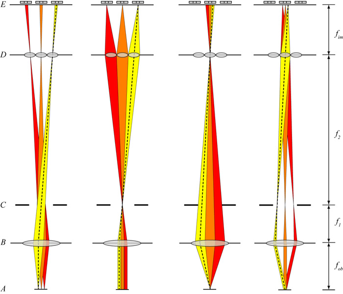 non-infinity optics, see memo for caption |
In our SIGGRAPH paper, figure 2 gives the optical layout of our prototype light
field microscope. Unfortunately, this figure and its accompanying caption do
not provide quite enough detail to set up and adjust a light field microscope.
This brief technical memo provides a more
detailed optical layout, with emphasis on how to vertically position and focus
each element in the optical train. We show this first for microscopes having
non-infinity-corrected optics (as in our SIGGRAPH paper). We then describe an
arrangement suitable for microscopes employing infinity-corrected optics, which
is the more common case. Finally, we address the often-asked question of
whether our microscope light fields contain aliasing.
(Yes they do.) |
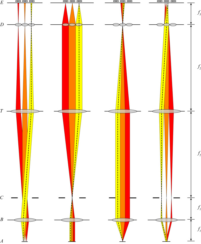 infinity optics |