|
|
|
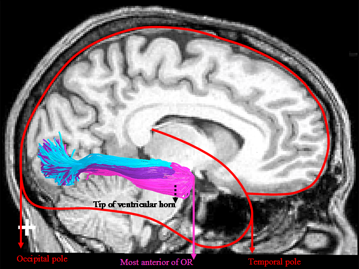
Figure 1. ConTrack estimates of the most likely pathways between the LGN and V1 match the gold-standard anatomy (Ebeling, 1988) in landmark distance estimates between the most anterior position of the optic radiation (OR) and 1) temporal pole, 2) tip of ventricular horn, and 3) occipital pole. As in Ebeling et al., the three sections of the OR have been labeled separately, the light purple labels the portion known as Meyer's loop. |
|
Measurement |
ConTrack X ± SD min-max (mm) |
Dissection (Ebeling) X ± SD min-max (mm) |
STT (Yamamoto) X ± SD min-max (mm) |
|
OR - Tip of temporal pole (distance) |
28 ± 3.0 |
27 ± 3.5 |
37 ± 2.5 |
|
OR - Tip of occipital pole |
96 ± 5.5 |
98 ± 6.2 |
82 ± 3.0 |
|
OR - Tip of ventricular horn (position) |
3 ± 2.6 |
5 ± 3.2 |
-4 ± 0.2 |
|
Table 1. ConTrack estimates on 16 hemispheres compared with the dissection of 25 hemispheres (Ebeling et al., 1988) and a previous DWI-tractography study of 5 hemispheres (Yamamoto, 2005). |