|
|
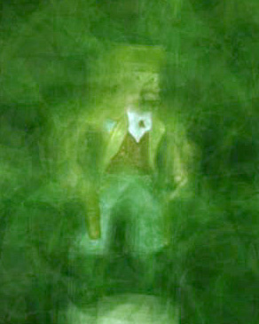
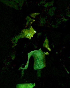
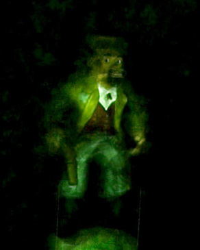
Using synthetic aperture photography and confocal imaging to see through
partially occluded environments. (a) shows one view of a figurine partially
obscured by a plant. Summing 16 different views produces an image (b) with a
wide synthetic aperture, hence a shallow depth of field, blurring out the
plant.
Alternatively, applying confocal imaging, by capturing a sequence of images
under patterned illumination as described in the paper, produces (c), in which
the plant has become dark. Combining these two techniques yields (d), in
which the plant is both blurry and dark, effectively disappearing.
|
|
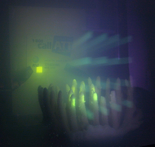
Using confocal imaging to enhance visibility in weakly scattering environments.
An AT&T calling card is placed in a tank filled with dilute milk. By
reflecting a video projector through an array of 16 mirrors, we create a
virtual projector having a synthetic aperture 1 meter wide. We use this
arrangement to illuminate the calling card with 16 converging beams (visible at
the right side of this photograph). We scan these beams across the card, and
for each beam position extract only those pixels inside a tile (bright
rectangle at left) where the beams intersect the card. Assembling these tiles
yields a composite image exhibiting less backscatter - hence better contrast -
than if the scene were floodlit. The results are in the paper.
|
Synthetic aperture confocal imaging
Proc. Siggraph 2004
Abstract:
Confocal microscopy is a family of imaging techniques that employ focused
patterned illumination and synchronized imaging to create cross-sectional views
of 3D biological specimens. In this paper, we adapt confocal imaging to
large-scale scenes by replacing the optical apertures used in microscopy with
arrays of real or virtual video projectors and cameras. Our prototype
implementation uses a video projector, a camera, and an array of mirrors.
Using this implementation, we explore confocal imaging of partially occluded
environments, such as foliage, and weakly scattering environments, such as
murky water. We demonstrate the ability to selectively image any plane in a
partially occluded environment, and to see further through murky water than is
otherwise possible. By thresholding the confocal images, we extract mattes
that can be used to selectively illuminate any plane in the scene.
Additional information available:
Confocal imaging versus separation of direct and global reflections
in 3D scenes
written by Marc Levoy
October 13, 2006
Many people have commented on the similarity between the enhanced underwater
visibility reported in our SIGGRAPH 2004 paper (linked to this web page) and
the removal of volumetric scattering effects reported in Nayar et al.'s
SIGGRAPH 2006 paper [1]. Even the discussions of illumination patterns are
similar in the two papers. However, the techniques proposed in the two papers
are different, as are their capabilities, and an analysis of these differences
is instructive.
The techniques described in our paper are based on confocal microscopy. In
particular, we describe two confocal imaging protocols. The first is based on
illumination of the scene by a scanned sequence of focused spots [2], and the
second on illumination of the scene by a sequence of focused random binary
patterns [3] followed by capture of one image under full illumination. A third
protocol that has been proposed in the microscopy literature is illumination of
the scene by a sequence of three sinusoidal patterns, shifted with respect to
one another by 1/3 of the pattern's wavelength [4,5]. In this protocol a
confocal image is formed by simple additions and subtractions of these three
images, without the necessity of extracting specific tiles.
We now make a number of observations about these protocols - observations that
we unfortunately overlooked when writing our SIGGRAPH paper:
-
The lack of a tile extraction step in the third protocol means that, although
the illumination source and camera are assumed to be focused at the same depth,
no particular lateral alignment needs to be assumed between the camera's lines
of sight and the projected patterns. This protocol is the basis for modern
structured-light microscopy systems [6].
-
Restricting ourselves to synthetic aperture confocal systems, in which focusing
is accomplished by an array of video projectors and cameras rather than by a
single optical aperture, and considering only the second protocol (random
binary patterns), although a minimum of two video projectors are required to
obtain confocal imaging when the camera is coaxial with one of the projectors
(figure 3f in our paper), only one projector is required in the non-coaxial
case (figure 3e). Indeed, in experiments conducted in a large water tank at
the Woods Hole Oceanographic Institution subsequent to writing the paper (see
below), we occasionally used only one projector - still
obtaining an improvement in visibility.
-
All three of these protocols perform optical sectioning, meaning that
they remove light reflected directly to the camera by points off the focal
plane. However, the third protocol (subtractions of shifted sinusoids) also
performs descattering. In particular, it removes light reflected to the
camera, whether by points on or off the focal plane, after multiple bounces.
By contrast, the scanning protocol removes none of this multiply-scattered
light, and the protocol based on random binary patterns removes some but not
all of it. A full analysis of the descattering ability of these various
protocols is beyond the scope of this technical note. Interestingly, this
author has been unable to find such an analysis in the confocal imaging
literature, where the assumption is usually made that the medium exhibits
minimal multiple scattering.
Now let's put these pieces together. Observation #1 says that confocal imaging
can be performed without calibration of the camera to the projector. Combining
this with observation #2, which says that one video projector suffices, means
that synthetic aperture confocal imaging can be applied to non-planar scenes,
in which no fixed relationship exists between the camera's lines of sight and
those of the projector. In those scenes the system will perform descattering
(as defined in observation #3) but not optical sectioning. In other words, it
can be used to remove global illumination effects from 3D scenes.
Unfortunately, we did not realize this in 2004. Nayar et al. did realize it,
although they approached the problem from the perspective of structured
illumination rangefinding rather than confocal imaging, and they therefore
expressed these observations differently than we would have. Applying these
ideas, they employed one projector and a protocol of three shifted sinusoids to
remove volumetric scattering effects from opaque non-planar objects immersed in
a weakly scattering medium (like the kitchen sink example in figure 6e of their
paper).
In summary, although both of these papers can be described in the language of
confocal imaging, the different choices of illumination protocols made by the
two papers led to very different capabilities. In particular, Nayar et al.'s
protocol based on a single projector and shifted sinusoids performs
descattering but no optical sectioning, whereas our protocol based on multiple
projectors and scanning or random binary patterns performs optical sectioning
but little or no descattering. If in our system we had used shifted sinusoids,
we could have performed both optical sectioning and descattering.
Note added June 16, 2008:
In a subsequent paper [7], we modified Nayar et al's technique to employ
multiple video projectors and cameras. This effectively combines his 2006
technique [1] and our 2004 technique (this web page), thereby performing both
optical sectioning and descattering of macroscopic scenes. Interested readers
are referred to the paper for details.
References
[1] Nayar, S.K., Krishnan, G., Grossberg, M.D., Raskar, R.,
Fast Separation of Direct and Global Components of a Scene
using High Frequency Illumination,
Proc. SIGGRAPH 2006.
[2] Corle, T.R.. Kino, G.S.
Confocal Scanning Optical Microscopy and Related Imaging Systems,
Academic Press, 1996.
[3] Wilson, T., Juskaitis, R., Neil, M., Kozubek, M.,
Confocal Microscopy by Aperture Correlation,
Optics Letters, Vol. 21, No. 3, 1996.
[4] Neil, M.A.A., Juskaitis, R., Wilson, T.,
Method of obtaining optical sectioning by using structured
light in a conventional microscope,
Optics Letters, Vol. 22, No. 24, 1997.
[5] Wilson, T., Neil, M.A.A., Juskaitis, R.,
Real-time three-dimensional imaging of macroscopic structures,
Journal of Microscopy,
Vol. 191, Issue 2, August 1998, pp. 113-220.
[6] Mitic, J., Anhut, T., Serov, Al, Lasser, T.,
Real-time optically sectioned wide-field microscopy
employing structured light illumination and a CMOS detector,
Three-dimensional and multidimensional microscopy:
Image acquisition and Processing X,
Proc. SPIE, Vol. 4964, January, 2003.
[7] Fuchs, C., Heinz, M., Levoy, M., Seidel, H.-P., Lensch, H.P.,
Combining Confocal Imaging and Descattering,
Eurographics Symposium on Rendering (EGSR) 2008.
Experiment in a large water tank at the
Woods Hole Oceanographic Institution

In our SIGGRAPH paper, we applied synthetic confocal imaging to two problems:
seeing through partially occluded environments such as foliage and crowds, and
seeing through turbid water. However, in the latter application we tested our
techniques only in a 10-gallon tank, with illumination and viewing distances of
10-30 cm. At that scale, backscatter of projected light to the camera
dominated over attenuation. In most practical applications of underwater
imaging, e.g. remotely operated vehicles (ROVs), illumination and imaging
distances are an order of magnitude larger, making attenuation more important.
Moreover, at the turbidities we employed in the 10-gallon tank, multiple
scattering was a significant factor, and as we point out in our paper, confocal
imaging performs poorly in the presence of multiple scattering. Finally, as we
explain in the note earlier on this web page about
confocal imaging versus separation of direct and global reflections, one
projector suffices to achieve confocal imaging if the projector is not
coaxial with the camera - a point we failed to emphasize in our paper.
To address these problems, in the Summer of 2004 (and consequently after we
published our SIGGRAPH paper), we repeated our underwater confocal imaging
experiment in a large water tank at the Woods Hole Oceanographic Institution
(WHOI). In this experiment, we used from 1 to 5 projectors. The "beauty shot"
reproduced above shows five projectors focusing a narrow beam (in green)
through turbid water (white haze) at a test target (not visible). Our most
important result from this experiment was that for oblique illumination from a
single projector, one obtains roughly an order of magnitude improvement in
SNR when imaging through turbid water if scanned confocal imaging is employed
instead of floodlighting. The experiment is described, and its results
analyzed, in this technical memo, co-authored with
Hanumant Singh of the WHOI Deep Submergence Laboratory. Although not described
in our SIGGRAPH paper, these results were presented during our SIGGRAPH 2004
talk, and they are included in the Powerpoint slide set linked above.
Copyright © 2004-2009 by Marc Levoy
|